ISTANBUL
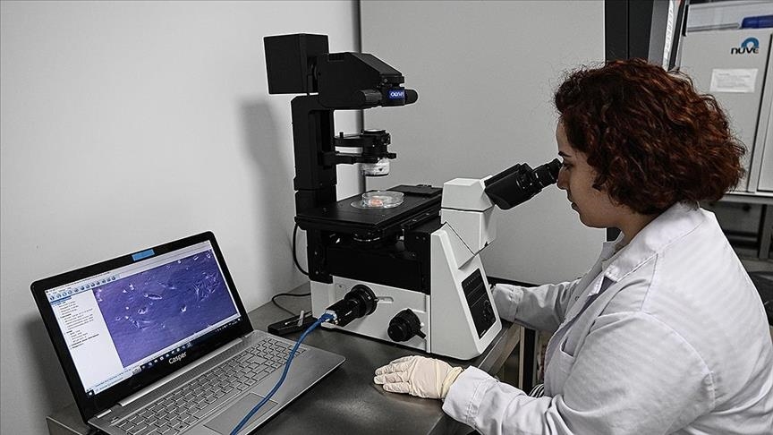
Working together, doctors and engineers from five Turkish universities have developed an imaging technique that will enable personalized treatment of cancer patients.
The scientists based their research on cell stiffness, which provides insight into the onset and progression of many different diseases, including cancer progression and differentiation.

Through some four years of studies, an imaging technique was implemented that will show the flexibility and stiffness of cells without the need to use chemicals, and give information about whether they are cancerous or not.
The technique, which could be used in many fields in the future such as parasitology, microbiology, mycology, blood diseases, food pathogens and pharmaceuticals, was published in the scientific journal Nature Communications.

Speaking to Anadolu Agency, Associate Professor Huseyin Uvet, the project manager, said cell architecture gives vital information for understanding many diseases.
“If we can make sense of the cell, it will mean developing various treatment modalities that can help us understand many diseases,” said Uvet.

“The cell may have a softer structure when it is cancerous or a harder structure when it is healthy. You can see this more clearly when it reaches the tissue level,” he explained.
“In structures such as breast cancer, you can see whether a tumor has formed by just touching it and looking at the hardness on the surface. We’re trying to capture this at the cellular level,” he added.

Uvet said they have gotten patents for the technique and are considering putting it on the market as microscopic imaging technology, adding: “A few years later, we plan to make it ready for use in hospitals and clinics where personalized treatment can be applied.”

